


Project objective
The sampling of tissues of brain tumors aiming at identifying the type of tumor and treatment planning is associated with significant problems and risks. On the one hand it is not clear which site on the tumor would be the best place to gain conclusive biopsy results with the effect that multiple tissue samples have to be taken. On the other hand there is a risk of causing cerebral hemorrhage when damaging a larger blood vessel while inserting the biopsy needle and also with each tissue sampling.
Funding announcement
Joint German-Russian funding competition of the Federal Ministry of Education and Research (BMBF) and the Russian Foundation for Assistance to Small Innovative Enterprises (FASIE) in the area of applied, industry-related research
Partner region / country
Russia
Life span
1 January 2015 – 30 June 2017
Partner institutions
MRC Systems GmbH, Heidelberg
Laser Research Center, Hospital of the Munich University
NeuroSpec LLC, Moscow
Burdenko Neurosurgery Institute, Moscow
Installing optical fibers into the biopsy needle shall solve these problems. The ideal place where tissue is removed is identified by marking it with a fluorescence dye. It has already proved successful in clinical use when surgically removing tumors. Ideally, a single sample of tissues is sufficient. With the aid of a spectral analysis of the light reflected by tissue, blood vessels should be identified in time in order to avoid cerebral hemorrhage. Optical biopsy needles are to be integrated into a mechanic target system. The signals should be illustrated in an imaging planning system along with computer tomography and magnetic resonance imaging.
Use of results
The medical device under development – the optical biopsy needle – as well as the associated hardware and software components is intended for neurosurgery, especially on suspicion of the presence of a malignant brain tumor. The existing Neurosurgery Centers in both partner countries will first benefit from the immediate market entry. There are 150 centers in Germany (www.dgnc.de) as well as 74 in the Russian Federation (www.rosminzdrav.ru). With a market share of 80%, about 180 units of this medical device could be realized.
In addition to this, it would be possible to manufacture similar devices using the same measuring principles to be used in other medical disciplines, e.g. for prostate and pancreatic tumors or breast carcinoma or for liver metastases.
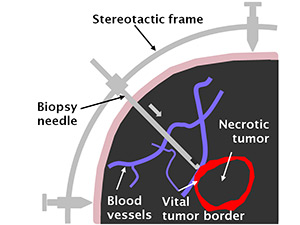
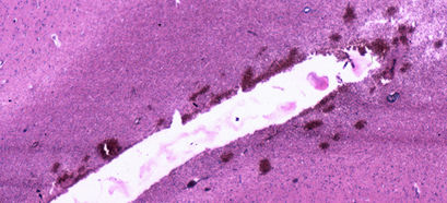
Tissue collection from brain tumors. A biopsy needle is attached to the stereotactic frame and inserted into the brain along a predefined direction (arrow) until the needle end is positioned in the vital edge (growth zone) of the tumor. The blood vessels (shown in blue) are recognized both in front of the biopsy needle and within the removal zone. Significant tissue samples must be taken from the vital tumor margin. This region is recognized by the red fluorescence of the selective fluorescence marker.
© Laser Research Center / Niklas Markwardt
Added value of international cooperation
The success of the project is only possible thanks to the cooperation of various partners. While the Russian industrial partner NeuroSpec contributes its experience and expertise in producing medical devices – specifically in opto-mechanical production –, the medical expertise of the Russian clinical partner Burdenko Neurosurgery Institute is indispensable for the user-friendly configuration of the product. The German academic partner Laser Research Center contributes its knowledge and decade-long experience in fluorescence diagnosis, tissue optics and fiber-based sensors, while the German industrial partner MRC Systems allows for the complete integration of the product including stereotactic target systems and software thanks to its expertise in medical and laser technology. After project completion medical approval in Europe will be pursued.
Outstanding results and achievements of the project
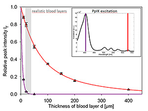
Attenuation of the fluorescence signal of the selective tumor marker PpIX by clinically relevant glow thicknesses at excitation with 405 nm (violet) and 633 nm (red). In spite of its low detection sensitivity when using the red excitation wavelength (small graphic), it is better suited because bleeding into the biopsy channel is much less disturbing.
© Laser Research Center / Niklas Markwardt
When pushing forward a biopsy needle into tissues with a good blood supply damaging small blood capillaries and flows of smaller amounts of blood are unavoidable. In this case excitation light in the red spectral range is better suited than the commonly used blue excitation light as this project was able to show.
Injuries to larger blood vessels have to be reliably avoided. A new spectroscopic method (dispersing radiation) was developed in order to recognize the injury under realistic conditions. This method can do without applying a contrast medium.
To date, four scientific publications (two in a peer-reviewed journal, two in conference proceedings) have been published. Moreover, a utility model was filed at the German Patent and Trade Mark Office.
DLR Project Management Agency
European and international Cooperation
Stefan Klumpp
phone: +49 228 3821-2038
MRC Systems GmbH
Innovative Medical and Laser Technology Products
Dr. Marcus Götz
phone: +49 6221 13803-13
